
FIGURE 6.
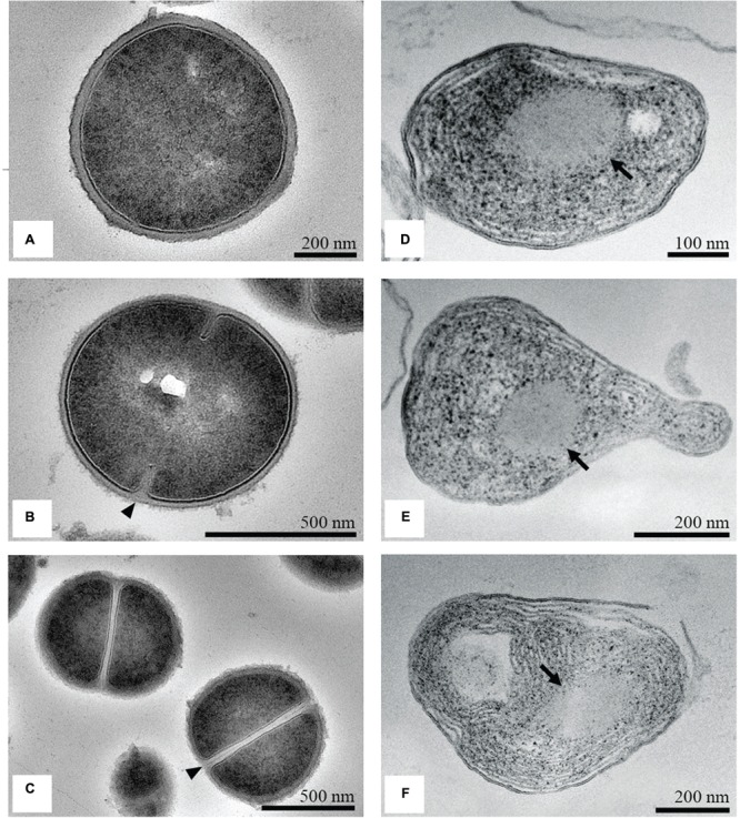
TEM micrographs of untreated and treated S. aureus. Untreated cells looked round and intact, with a well-defined cell membrane (A). The onset of septation (arrowhead; B) and the cross-wall formation (arrowhead; C) was noticed. After treatment with the MIC of the methanol extract of C. versicolor, cells appeared elongated and malformed, with non-membrane-enclosed bodies (denoted by arrows; D–F). Some lysed cells were also noted (F).