
FIGURE 7.
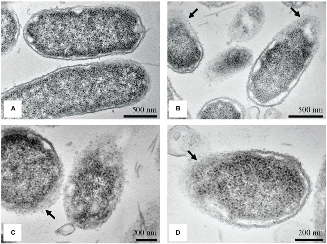
TEM micrographs of untreated and treated S. Enteritidis. A regular outlined cell envelope and regularly distributed cytoplasm lying closely to the cell wall were observed with untreated cells (A). After treatment with the MIC of the methanol extract of C. versicolor, unsymmetrically distributed cytoplasm, larger and irregular periplasmic space and deformed and scattered components of the cell envelop were observed (B–D). The cells without the membranes (C) as well as null cells (D) were also noted.