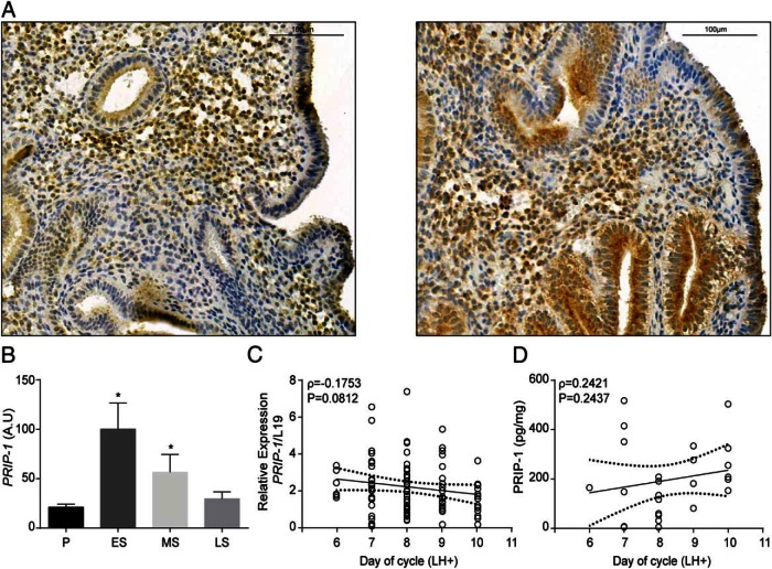
Figure 2. PRIP-1 expression in midluteal endometrium.
A, Immunohistochemistry of midluteal endometrial biopsies demonstrates heterogeneous expression of PRIP-1 in both stromal and epithelial cells. B, Comparison of endometrial PRIP-1 transcripts, expressed in arbitrary units (A.U.), in proliferative, early-, mid-, and late-secretory endometrium. The data were derived from in silico analysis of publicly available microarray data (GEO Profiles; ID, GDS2052); *, P < .05. C, PRIP-1 transcript levels were measured by qRT-PCR in 73 endometrial biopsies obtained between 6 and 10 days after the LH surge (LH+). D, PRIP-1 protein levels were measured, in triplicate, in 25 whole endometrial samples by ELISA. Dotted lines represent 95% confidence intervals.

