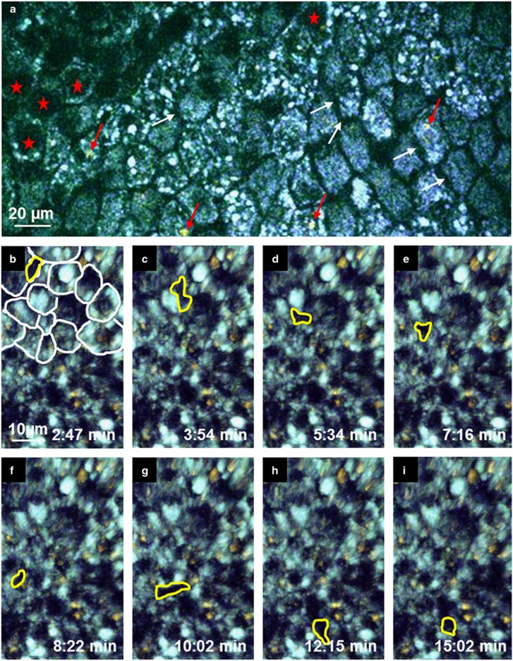
Figure 3.
Cell dynamics in the tracheal epithelium. En face view of the epithelium. (a) Epithelial cells were identifiable by autofluorescence singal surrounding a non-fluorescent nucleus (red asterisks). The borders between the cells were non-fluorescent (white arrows). In some cells, a red shifted autofluorescence signal (red arrows) was observed. (b–i) The Image series shows a non-fluorescent motile cell (framed by a yellow line) moving through the basal part of the epithelium (part of the epithelial cells were surrounded by white lines) by pushing epithelial cells to the side (see Supplementary Movie 4).

