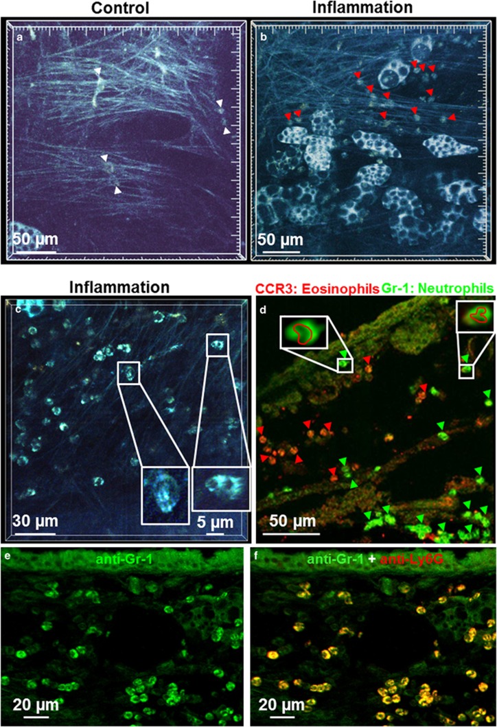
Figure 5.
Inflammatory cells in airway inflammation visualized by multiphoton microscopy. (a and b) Time series of a projection of 10 focal planes (a) and 9 focal planes (b). (a) In the connective trachea of control animals, very few cells were observed (white arrowheads (Supplementary Movie 11). (b) During an acute airway inflammation, a large number of motile cells were visible (red arrowheads; see also Supplementary Movie 11). (c) These cells showed a typical morphology with a lobed nucleus (see inset with magnification of boxed area in c). (d) Immunohistochemical staining of a cryostat section from a specimen previously examined by multiphoton microscopy identified these cells as CCR3-immunoreactive eosinophils (red, red arrowheads) and GR-1-immunoreactive neutrophils (green, green arrowheads). (e and f) Double labeling with anti-GR-1 antibody (green) and anti-Ly6G antibody (red) confirmed that GR-1 staining identifies neutrophils (appearing yellow in f).

