Figure 7.
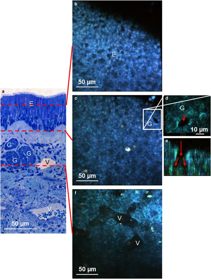
Imaging of human nasal concha ex vivo using multiphoton microscopy. (a) Semithin section of nasal concha stained with methylene blue-azure II. Red dashed lines indicate the approximate depth where the en face multiphoton images were taken. (b) En face view of the autofluorescent epithelium (E). (c) En face view of the connective tissue directly under the epithelium. G, gland. (d) Magnification of boxed area in (c) red arrow labels opening of gland. (e) Cross-section through z-stack showing the bifurcation of the gland. (f) Connective tissue 20 μm under the epithelium with vessels (V). For the complete z-stack see Supplementary Movie 15.

