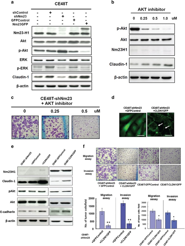
Figure 4.
Nm23H1 expression regulates Akt phosphorylation and CLDN1 expression in the CE48T cells, and expression of CLDN1GFP in the CE48T cells suppresses cell migration and invasion independently of Nm23H1. (a) Western blot analyses of Nm23H1, Akt, p-Akt, ERK, p-ERK, CLDN1 and β-actin in the shRNA-control (shRNA vector), GFP control (GFP vector), Nm23H1-silencing (shNm23), Nm23H1-GFP and parental CE48T cells were shown. (b) Western blot analysis of p-Akt, Akt, Nm23H1, CLDN1 and β-actin in the CE48T-shNm23 cells treated with the AKT inhibitor (MK2206) at the indicated concentration. (c) Effects of MK2206 on migration of the CE48T-shNm23 cells were assessed by Transwell migration assay. Original magnification, × 100. (d) CLDN1GFP was predominantly localized to the membrane of the Nm23H1-silencing cells at cell–cell contact points (arrows), a pattern that was morphologically similar to the TJ strand network in situ, whereas a diffuse cytosolic GFP signal was found in the GFP vector-transfected Nm23H1-silencing cells. (e) Western blot analysis for the protein level of CLDN1, pAkt, Akt, E-cadherin and β-actin in the CE48T-shNm23 cells and the CE48T cells with or without CLDN1GFP expression. (f) The migration and invasion assays of the representative results were shown. The bar graphs presented the mean values obtained from three independent determinations (**P<0.01; *P<0.05).

