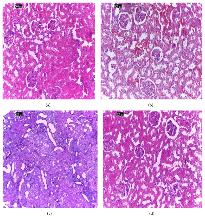
Figure 1.
(a) Photomicrography of a kidney of negative control group (G1) reveals normal histological structure; (b) photomicrography of a kidney of the positive control group with pathological changes; (c) photomicrography of a kidney of G3 treated with L. sativum shows nearly normal tissues; (d) photomicrography of a kidney of G4 group treated with cinnamon shows nearly normal renal cortical tissue (H&E ×200).

