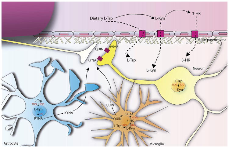
Fig. 3. The Kynurenine pathway in the brain.
Brain kynurenines are linked and influenced by the peripheral KP. Fluctuations in the blood levels of L-Trp, L-Kyn and 3-HK directly affect metabolism of KP in the brain, since these metabolites readily cross the BBB using the large neutral amino acid transporter. Under physiological conditions, kynurenine pathway enzymes in the mammalian brain are preferentially, although not exclusively, localized in non-neuronal cells (see below) and the two routes of L-Kyn degradation are physically segregated in the brain. In fact, astrocytes, which contain KAT, are the main responsible of kynurenic acid (KYNA) biosynthesis, a well established NMDA receptor antagonist. On the contrary, microglial cells generate 3-HK and its major downstream metabolites, including quinolinic acid (QUIN), which present neurotoxic properties, at least in part, by being an NMDAR agonist. Both QUIN and KYNA are released into the extracellular space and act over NMDA receptors in the postsynaptic compartment of the neurons. In addition, under several conditions, neurons can also express different kynurenine pathway enzymes, such as TDO and IDO.

