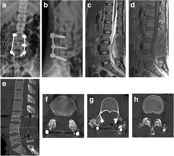
Fig. 3.

The postoperative X-rays and CT and MRI scans. (a-b) Images of postoperative X-rays. Sagittal T2WI (c) and T1WI (d) of the postoperative MRI images show the mass has been completely resected. (e-h) Images of postoperative CT confirm that the mass has been removed. e shows the sagittal plain and f-h represent the level of L3, L4 and L5, respectively
