
Fig. 1.
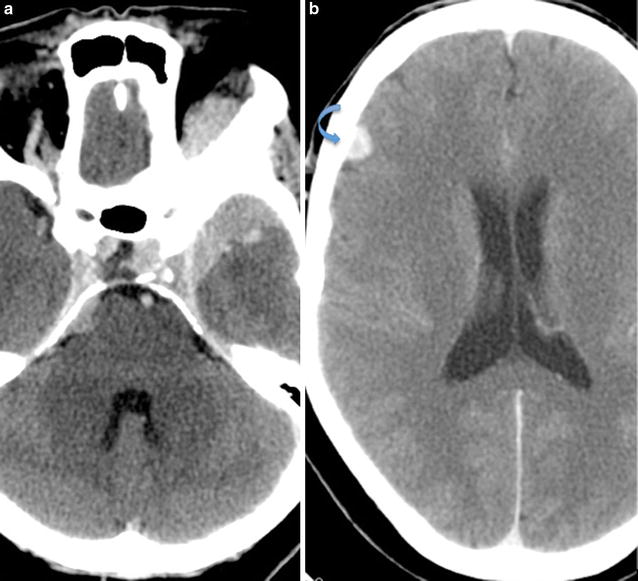
Axial contrast enhanced CT of the brain. a Large homogenously enhancing left sphenoid wing meningioma with associated hyperostosis and extension into the left anterior temporal fossa and left extraconal space. b Smaller well-defined enhancing extra-axial lesion in the right frontal lobe (arrow), consistent with a convexity meningioma