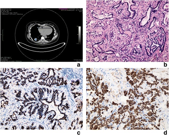
Fig. 1.

Lung tumour: Metastatic adenomyoepithelioma with component of epithelial-myoepithelial carcinoma. a PET-CT scan: Two nodular masses located in right lower (measuring 26x31mm) and middle lobe (diameter 96 mm). b Light microscope. Inner layer of glandular structures lined by epithelial cells, outer layer formed by myoepithelial cells with clear cytoplasm, mild nuclear atypia and some nucleoli visible. Homogenous, hyalinised stroma. H + E x400. c Immunochistochemical staining for cytokeratin positive in epithelial cells. x400. d Immunochistochemical staining for SMA positive in myoepithelial cells. x400
