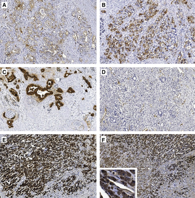
Figure 3.
Representative pancreatic tumour tissue samples at × 10 magnification. (A) Intermediate and (B) strong TF staining in moderately and poorly differentiated pancreatic adenocarcinoma, respectively, (C) MUC1 staining, (D) CD31 staining of endothelium, (E) CD68 staining of large clusters of macrophages and (F) corresponding very strongly TF+ macrophages.

