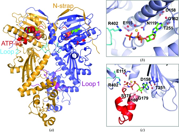
Figure 1.
Crystal structure of human TRAP1NM bound to ADPNP. (a) Ribbon diagram depicting the TRAP1NM dimer with ADPNP shown as a CPK model. Each subunit is colored differently. Key structural elements are labeled. (b) Enlarged view of the N-terminal ATP-binding pocket of one TRAP1NM subunit with bound ADPNP (stick model). Ordered water molecules are shown as red spheres and the bound Mg2+ ion as a yellow sphere. (c) Enlarged view of the bound ADPNP molecule with the ATP-lid colored red.

