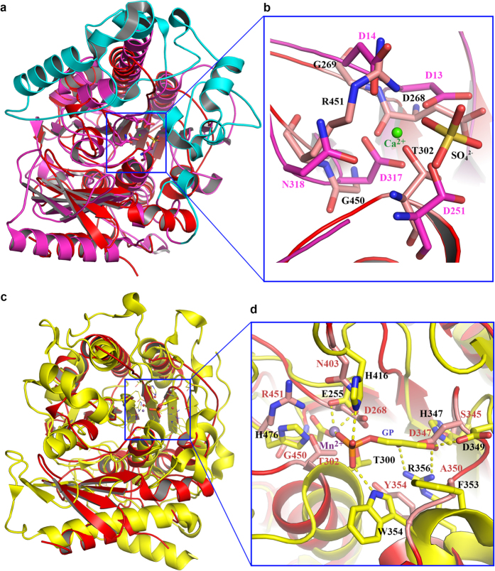
Figure 3. PbgA structure is similar to arylsulfatase and lipoteichoic acid synthase, but do not contain the enzyme catalytic residues.
(a) StPbgA (red) is superimposed to arylsulfatase (matanga). The arylsulfatase has additional α-helices and loops (cyan), forming the substrate binding “gate”. (b) Superimposition of StPbgA and arylsulfatase at the active site. (c) StPbgA (red) is superimposed to lipoteichoic acid synthase (yellow). (d) Superimposition of StPbgA and lipoteichoic acid synthase at the active site.

