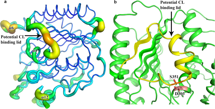
Figure 5. Potential CL binding lid.
(a) The PbgA loop consisting of residues D347-Q370 has higher B-factor comparing to the local residues, suggesting that the loop is flexible and may be a lid for the CL binding. (b) Cartoon representation of potential CL binding lid colored in yellow and the double cysteine mutation residues S351C/D303C may form a disulphide bond and prevent the lid to open for CL binding.

