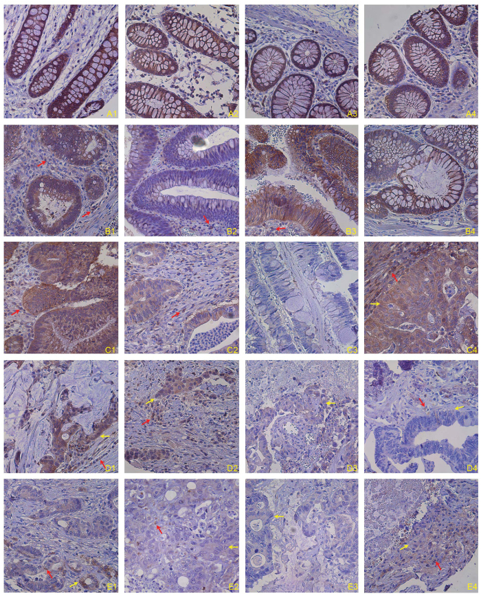
Figure 1. Fas protein level in normal human colon and human colorectal carcinoma tissues.
Human colon carcinoma tissues were stained with anti-human Fas monoclonal antibody. Brown color indicates Fas protein level, with counterstaining by hematoxylin in blue. Shown are representative images of adjacent normal human colon tissues from colon cancer patients (A1–4 indicates tissues from four patients), adenomas (B1–4), primary invasive adenocarcinoma (C1–4), colorectal adenocarcinoma metastatic to lymph nodes (D1–4), and colorectal adenocarcinoma metastatic to liver (E1–4).

