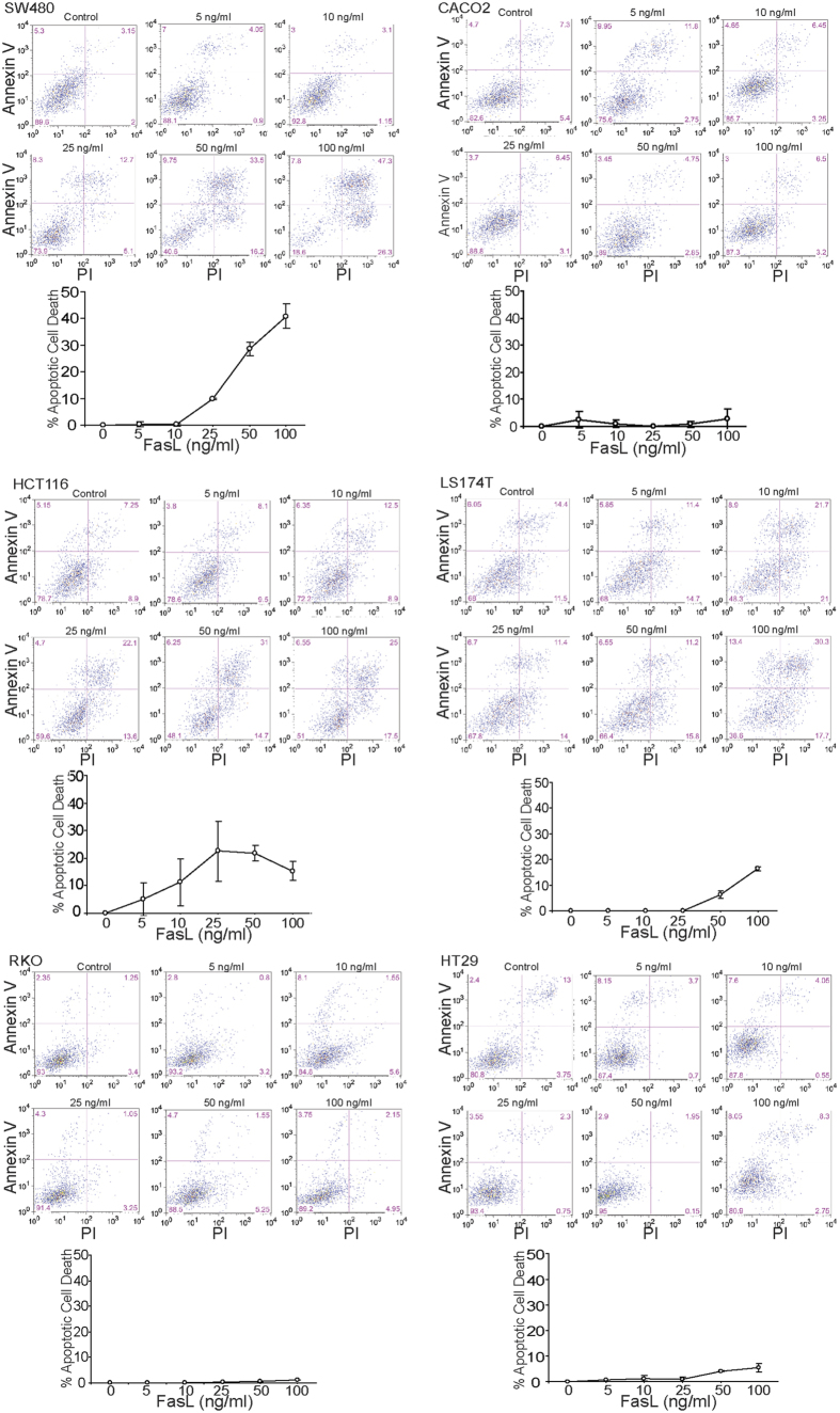
Figure 2. Sensitivity of human colon carcinoma cells to FasL-induced apoptosis.
The indicated human colon carcinoma cells were cultured in the presence of MegaFasL at the indicated concentrations for approximately 24 h. Both floating and adherent cells were harvested and stained for Annexin V and PI. Cells were analyzed by flow cytometry. For each cell line, the top panel shows representative plots of apoptotic cell death. The bottom panel shows quantification of apoptotic cell death. Percent apoptotic cell death was calculated as (% Annexin V+PI+ cells in the presence of FasL) − (% Annexin V+PI+ cells in the absence of FasL). Column: mean; Bar:SD.

