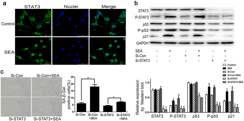
Figure 5. SEA-induced LX-2 senescence is dependent on STAT3.
(a) The expression of STAT3 in LX-2 cells was determined by immunocytochemistry using a confocal laser scanning microscope, and the nuclei were stained with Hoechst 33342. (b) Western blot analysis for STAT3, P-STAT3 (Y705), p53, P-p53 (S15) and p21 proteins in Si-STAT3-treated LX-2 cells. *p < 0.05 compared to the control group; &p < 0.05 compared to the Si-Con group; $p < 0.05 compared to the control group; #p > 0.05 compared to the Si-STAT3 group. (c) A representative image from SA-β-Gal staining and percentage (%) of SA-β-Gal -positive cells following Si-Con or Si-STAT3 treatment with or without SEA. Bar: 50 micrometers. Statistical differences between groups are shown as follows: *p < 0.05; **p < 0.01.

