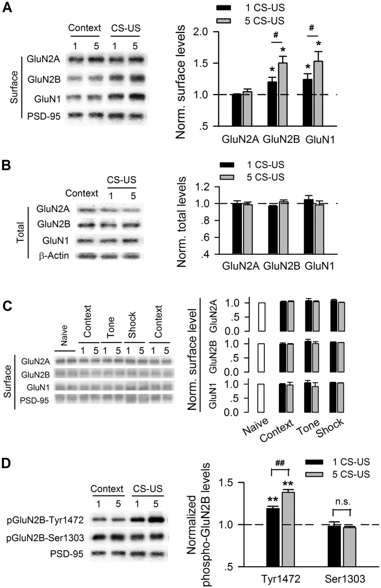
Figure 2. The amount and phosphorylation of membrane GluN2B-containing NMDARs exhibit a conditioning-strength-dependent increase after fear conditioning.
(A) Immunoblots (left) and quantification analysis (right) of surface NMDAR subtypes in membrane lysates of CA1 taken from rats unconditioned (context) versus rats conditioned with single (1 CS-US) or five CS-US (5 CS-US), normalized to context control. (B) Immunoblots (left) and quantification analysis (right) of NMDAR subtypes in total lysates of CA1 taken from rats with the same treatments as in (A). (C) Immunoblots (left) and quantification analysis (right) of surface NMDAR subtypes in membrane lysates of CA1 taken from naïve, context alone, tone alone, and shock alone rats, normalized to naïve rats. (D) Immunoblots (left) and quantification analysis (right) of GluN2B phosphorylation at Tyr1472 and Ser1303 in membrane lysates of CA1 taken from rats with the same treatments as in (A). *p < 0.05, **p < 0.01 vs. context-control; #p < 0.05, ##p < 0.01.

