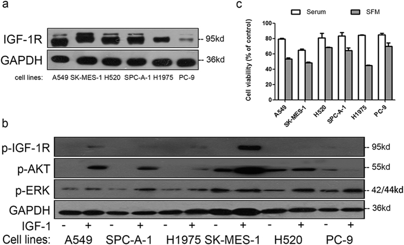
Figure 1. IGF-1R expression and sensitivity to CP treatment in NSCLC cell lines.
(a) NSCLC cell lysates were prepared and IGF-1R were detected by WB. (b) Cells were starved for 12h and stimulated with 50 ng/ml IGF-1 for 10 min. p-IGF-1R, p-ERK, p-AKT and GAPDH were detected by WB. (c) Cells were treated with 100 ng/ml of CP for 48 h with presence or absence of serum. Cell viability was tested through MTT. The number of viable cells following CP treatment was presented as percentage of untreated cells. Data were presented as mean ± S.E.M.

