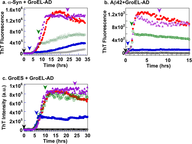
Figure 5. Delayed addition of GroEL-AD to the fibril forming reactions of each client protein.
In each panel, colored arrowheads denote the instant at which excess GroEL-AD was added to each corresponding color-coded trace of the experiment. (a) α-Synuclein. GroEL-AD (3-fold molar excess) was added at 0 (black), 3 (blue), 8 (green) and 24 (magenta) hours after initiating the experiment. (b) Aβ42 peptide. GroEL-AD (20-fold molar excess) was added at 0 (black), 0.5 (blue), 1.5 (green) and 8 (magenta) hours after initiating the experiment. (c) GroES in 0.4 M Gdn-HCl. GroEL-AD (4-fold molar excess) was added at 0 (black), 6 (blue), 10 (green) and 24 (magenta) hours after initiating the experiment.

