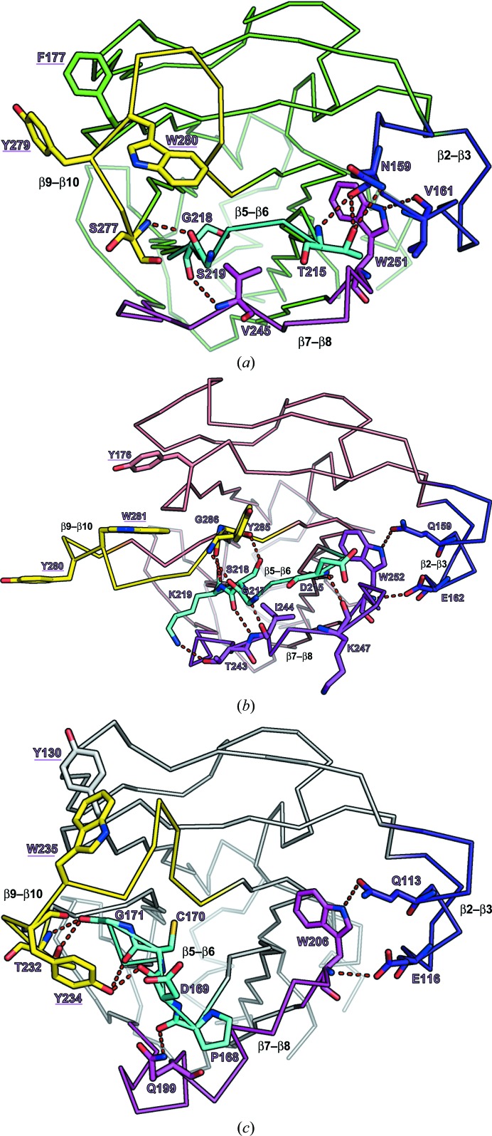
Figure 4.
Structures of the substrate-binding pockets of wild-type Fbs1 SBD (a), loop-mutant 1 (b) and the FBG3 SBD (c). Hydrogen bonds are represented as dashed red lines. Residues of hydrogen-bonding pairs and the carbohydrate-binding pocket are depicted as stick models. The four loops β2–β3, β5–β6, β7–β8 and β9–β10 are coloured blue, cyan, magenta and yellow, respectively.

