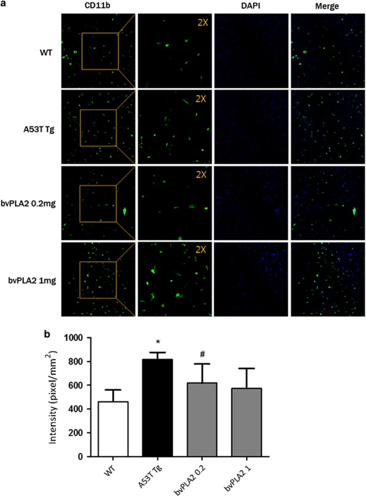
Figure 3.
Treatment with bvPLA2 induces microglial deactivation in the spinal cord. (a) A section was selected and processed for CD11b immunofluorescence. Confocal microscopic observation revealed microglia fluorescence (green) in the spinal cord. The nuclei were counterstained with DAPI (blue). The insets are × 2 magnification of the boxed area. The representative immunostained α-Syn inclusions are indicated as arrows. (b) The confocal image shown in (a) was analyzed. The values indicate the mean±s.e.m. *P<0.05 compared with the WT group, #P<0.05 compared with the A53T Tg group. α-Syn, α-Synuclein; Tg, transgenic; WT, wild type.

