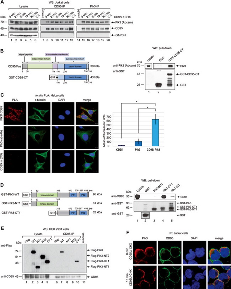
Figure 1.
Plk3 interacts with CD95. (A) Jurkat cells were treated with CD95L and CHX for different time periods. CD95 or Plk3 was immunoprecipitated from cell lysates with anti-CD95 mouse or anti-Plk3 rabbit (Abcam) antibodies. Normal mouse or rabbit IgG was used as a control. The precipitated proteins were immunoblotted for Plk3 and CD95. The Plk3 (Abcam) antibody recognized two bands corresponding to the information by the manufacturer. (B) Schematic representation of the CD95/Fas receptor and the GST-fused cytoplasmic C-terminal subdomain of CD95 (GST-CD95-CT) (left panel). Binding of GST-CD95-CT with endogenous Plk3 was analyzed in pull-down assays. GST-CD95-CT or GST alone (control) was incubated with the lysates of HeLa cells (right panel). Endogenous Plk3 was detected by immunoblotting. (C) The interaction of Plk3 and CD95 was monitored via in situ Proximity Ligation Assay (PLA). HeLa cells were labeled with anti-Plk3 (rabbit) and anti-CD95 (mouse) antibodies. Single antibody staining (Plk3/CD95) was used as a control. Scale bar: 10 μm. The quantification is shown in the right panel. Differences between single and double antibody staining were statistically significant by Student's t-test (*P ≤ 0.05). (D) Schematic representation of recombinant GST-fused full-length Plk3 (GST-Plk3-WT) and truncated forms of Plk3 (GST-Plk3-NT1 and -CT1) (left panel) used in pull-down assays. GST-Plk3-WT or Plk3 subdomains were incubated with Jurkat cell lysates for pull-down assays. Endogenous CD95 was detected by immunoblotting. (E) HEK 293T cells were transfected with Flag-Plk3-WT and its truncated forms (Supplementary information, Figure S2D) for 24 h. Endogenous CD95 was immunoprecipitated from cell lysates with anti-CD95 mouse antibody. Normal mouse IgG was used as a control. The precipitated proteins were immunoblotted for CD95 and Flag. (F) Co-localization of Plk3 and CD95 with or without CD95L treatment was studied using immunofluorescence microscopy. Cells were stained for Plk3, CD95 and DNA. Representative image acquisition was performed using a confocal laser-scanning microscope (CLSM) and rendering of confocal z stacks was performed using the LAS AF software. Scale bar: 2 μm.

