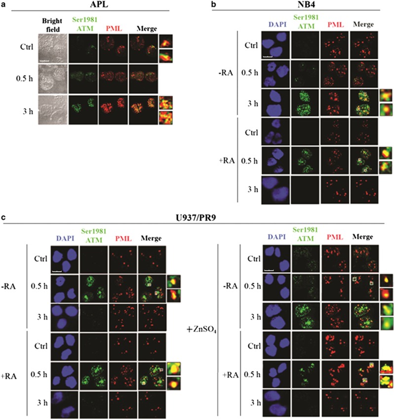
Figure 5.
PML-NB integrity and ATM activation. Double immunofluorescence of pSer1981-ATM (Alexa Fluor 488, green fluorophore) and PML (Alexa Fluor 610, red fluorophore) foci performed in cells untreated or irradiated with 1 Gy and fixed after 0.5 and 3 h. Representative images of the analysis performed in (a) APL blasts (cell image: bright field), (b) NB4 cells untreated or treated with 1 μM RA for 72 h previous to IR (counterstain: DAPI), and (c) U937/PR9 cells either untreated or exposed for 8 h to 100 μM ZnSO4 and then untreated or treated with 1 μM RA for 72 h previous to IR (counterstain: DAPI). Some colocalization signals have been highlighted and marked within the cell by a white square. Confocal microscopy images, magnification × 63; LCS Leica confocal microscope (Leica Microsystems)

