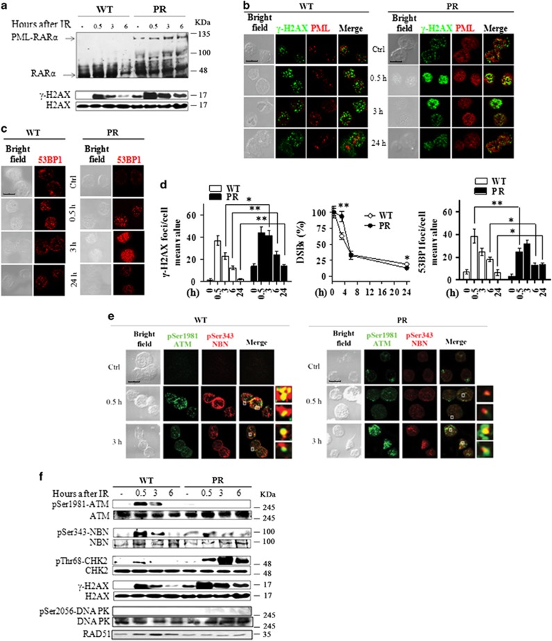
Figure 8.
In vivo validation of the DDR in the preleukemic mouse model of APL. WT and preleukemic mice knock-in for PML-RARα (PR) were irradiated with 5.5 Gy of X-rays and sacrificied after 0.5, 3, 6, and 24 h. Lin− cells were isolated from the bone marrow of three pooled mice. (a) Immunoblot analysis of RARα and PML-RARα expression, and of H2AX phosphorylation at Ser139 residue, in untreated and irradiated WT and PR mice. (b) Representative images of the double immunofluorescence analysis of γ-H2AX (Alexa Fluor 488, green fluorophore) and PML (Alexa Fluor 610, red fluorophore) foci in untreated and irradiated WT and PR mice. (c) Representative images of the 53BP1 foci in untreated and irradiated WT and PR mice. (d) The DSBs rejoining analysis was reported as the mean value of γ-H2AX foci/cell and as the percentage of residual DSBs in untreated and irradiated WT and PR mice. The DSBS repair was also analyzed by counting the number of 53BP1 foci/cell in untreated and irradiated WT and PR mice. Mean values were derived from the analysis of 100 cells from three independent experiments±S.D. *P<0.05, **P<0.01. (e) Representative images of the double immunofluorescence analysis of pSer1981-ATM (Alexa Fluor 488, green fluorophore) and pSer343-NBN (Alexa Fluor 610, red fluorophore) foci in WT and PR mice. (f) Immunoblot analysis of ATM phosphorylation at the Ser1981 residue, NBN phosphorylation at Ser343, CHK2 phosphorylation at Thr68, DNA-PK phosphorylation at Ser2056, and RAD51 expression. Cell image: bright field; confocal microscopy images, magnification × 63; LCS Leica confocal microscope (Leica Microsystems)

