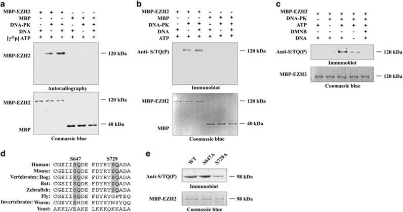
Figure 4.
DNA-PKcs phosphorylates EZH2 in vitro. (a) An in vitro reaction system was set up in 50 μl of reaction buffer containing 20 units of DNA-PK complex, 200 μg MBP-EZH2, 200 μM ATP, 5 μCi (γ-32P) ATP, 4 μg BSA, and 10 μg/ml calf thymus DNA. The mixture was collected after a 15 min incubation at 37 °C, the signal was detected by autoradiography, and a parallel gel was stained with Coomassie blue. (b) ATP without (γ-32P) was added to the reaction mixture. EZH2 phosphorylation was analyzed by immunoblotting and Coomassie blue staining. (c) DMNB (5 μM) was added to the reaction mixture. EZH2 phosphorylation was analyzed by immunoblotting and Coomassie blue staining. (d) Schematic alignment of EZH2 S/TQ sites in different species. (e) S647A and S729A mutants were generated and subjected to kinase assays. EZH2 phosphorylation was analyzed by immunoblotting and Coomassie blue staining

