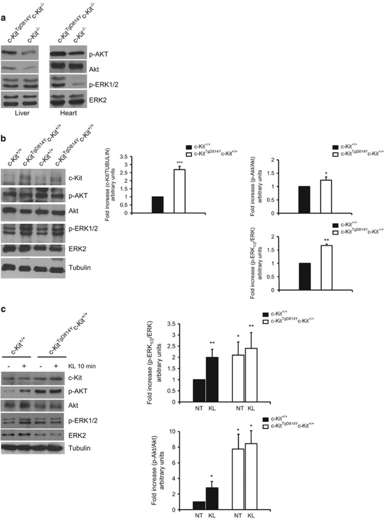
Figure 2.
MAPK and AKT activation in c-KitTgD814Y transgenic hearts. (a) Hearts and livers from c-KitTgD814Yc-Kit−/− and c-Kit−/− E15.5 were isolated and protein extracts analyzed for AKT and MAPK phosphorylation. Phosphorylated substrates were increased in embryonic hearts from transgenic mice. (b) WB analysis on protein extracts from c-KitTgD814Yc-Kit+/+ and c-Kit+/+ 3 dpp hearts of two different animals. Densitometry of c-Kit expression, MAPK and AKT phosphorylation is reported. Data are reported as mean of three hearts±S.D.; *P<0.05, **P<0.01, ***P<0.001. (c) Neonatal cardiac cells isolated from c-KitTgD814Yc-Kit+/+ and c-Kit+/+ 3 dpp hearts were stimulated with 100 ng/ml of KL for 10 min and protein extracts processed for AKT and MAPK phosphorylation. Densitometry of six separate experiments is reported. Data are reported±S.D.; *P<0.05, **P<0.01

