Figure 4.
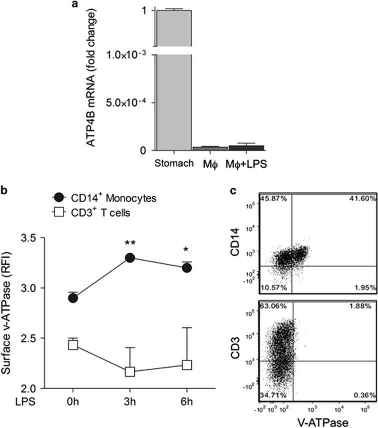
Macrophages do not express the gastric H+/K+ ATPase, but display surface v-ATPases. (a) Reverse transcription-polymerase chain reaction (RT-PCR) analysis of mRNA coding for the β-subunit of the gastric H+/K+ proton pump on peritoneal mouse macrophages (Mφ) untreated or exposed 3 h to LPS (Mφ+LPS). Data are expressed as fold changes of normalized expression versus murine stomach (mean±S.E.M. of three experiments). (b and c) PBMCs from healthy donors were double stained with anti-v-ATPase and anti-CD14 antibody (Ab) or anti-CD3 Ab time 0 or 3 or 6 h after exposure to LPS and analyzed by FACS. In (b), data are expressed as the relative fluorescence intensity (RFI) of v-ATPase in CD14+ (monocytes) and CD3+ (T-lymphocytes) cells (mean±S.E.M. of three independent experiments). Statistical analysis is referred to monocytes and evaluated versus t0. *P<0.05; **P<0.01 versus (c) a representative experiment of costaining (out of 3) is shown: 41% of CD14+ cells (upper plot) and 1.88% of CD3+ cells are positive for surface v-ATPases at 3 h from LPS exposure. *P<0.05; **P<0.01

