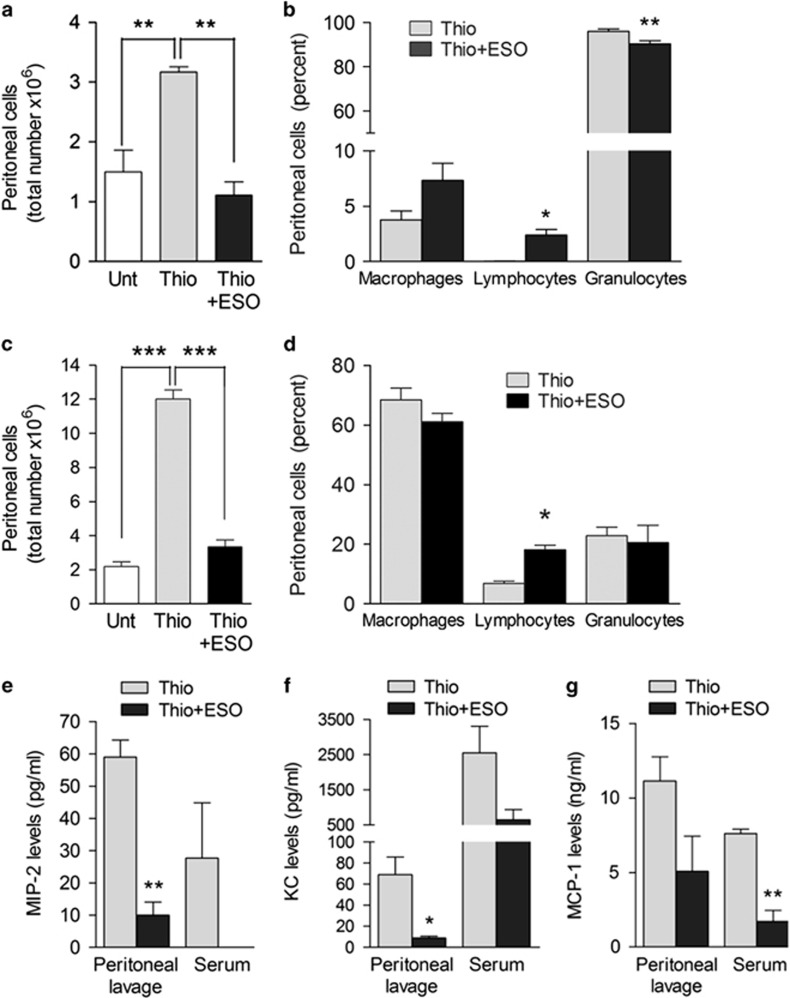
Figure 8.
ESO prevents thioglycollate-induced peritonitis. (a and c) Peritoneal cells isolated 4 h (a) or 72 h (c) from untreated (Unt) mice or from mice injected intraperitoneally with thioglycollate alone (Thio) or thioglycollate 30 min after intraperitoneal injection with ESO (Thio+ESO) were counted. Data are expressed as the total number of infiltrating inflammatory cells (mean±S.E.M.; N=3). (b and d) The relative percent of macrophages, lymphocytes and granulocytes in peritoneum lavage from the same mice was calculated at 4 h (b) and 72 h (d). (e, f and g) MIP-2 (e), KC (f) and MCP-1 (g) levels were determined in serum and peritoneal lavage 4 h after thioglicollate treatment in untreated or ESO-treated mice (pg/ml; N=3, mean±S.E.M.) *P<0.05; **P<0.01; ***P<0.001

