Figure 5.
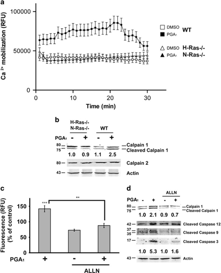
PGA1 induces calpain activation. Serum-starved wt or H-Ras−/−/N-Ras−/− MEFs were stimulated with 30 μM PGA1 or DMSO. (a) Increase in cytosolic Ca2+ levels. Measurements are given in relative fluorescent units (RFU) during 30 min. Data are expressed as mean±S.D. of three independent experiments. (b) Cell lysates were obtained 3 h after treatment and analyzed for detection of calpain-1, calpain-2, and actin. (c) Calpain activity was measured 3 h after treatment. Measurements are given in RFUs. Data are expressed as mean±S.D. (n=3). ***P≤0.001 versus control and **P≤0.01. (d) Serum-starved wt MEFs were stimulated with 30 μM PGA1 or DMSO in the presence of the calpain inhibitor ALLN (10 μM). Cell lysates were obtained 3 h after treatment and analyzed as in Figure 2b and in (b). Levels of cleaved calpain-1 and caspase-3 are denoted at the bottom of each panel (S.D. <10% average in each case, n=3)

