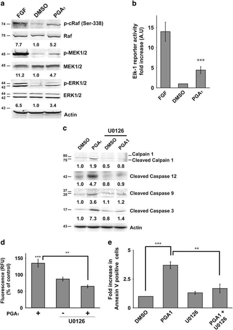
Figure 6.
ERK activation is necessary for PGA1-dependent apoptosis. Serum- starved wt MEFs were stimulated with 30 μM PGA1, FGF, or DMSO. (a) Lysates prepared from cells 15 min after treatment underwent western blotting for relevant components of the RAF-MEK-ERK pathway. Levels of p-c-RAF, p-MEK1/2, and p-ERK1/2 are provided at the bottom of each panel (S.D. <10% average in each case, n=4). (b) Cells were co-transfected with the plasmids pcDNAIII-Gal4-Elk-1, pGal4-Luc, and pTK-Renilla 24 h before synchronization. Firefly luciferase activity was determined 6 h after stimulation with PGA1 and normalized to Renilla luciferase activity. Results, in arbitrary units (AU), are expressed as mean±S.D. (n=3). ***P≤0.001 versus control. (c) MEK1/2 inhibitor U0126 (5 μM) was added ~50 min before PGA1 treatment (30 μM) and left in the medium until processing. Cell lysates were prepared and analyzed as in Figure 5c. Levels of cleaved calpain-1, cleaved caspase-12, cleaved caspase-9 and caspase-3 are indicated at the bottom of each panel (S.D. <10% average in each case, n=3). (d) Calpain activity was measured as in Figure 5d. Data are expressed as mean±S.D. (n=3). ***P≤0.001 versus control and **P≤0.01. (e) Annexin V/FITC staining analysis was performed as in Figure 1c. Results are expressed mean±S.D. (n=4). ***P≤0.001 and **P≤0.01

