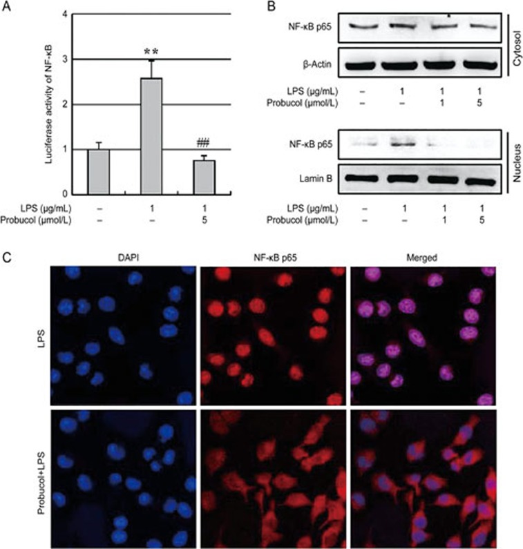
Figure 3.
Effect of probucol on NF-κB activity. (A) Transfected 293-T cells were pretreated with 5 μmol/L probucol for 3 h and then stimulated with LPS (1 μg/mL) for 6 h. NF-κB activity is expressed as the relative luciferase activity. The results are expressed as the mean±SEM (n=3). **P<0.01 vs cells without LPS; ##P<0.01 vs cells treated with LPS in the absence of probucol. (B) The total cytosolic and nuclear proteins were subjected to Western blotting using anti-NF-κB p65. Actin and lamin B were used as internal controls. (C) The localization of NF-κB p65 was visualized by confocal microscopy following immunofluorescence staining with anti-NF-κB p65 (red). The cells were stained with DAPI to visualize the nuclei (blue). The results are representative of those obtained from three independent experiments.

