Figure 7.
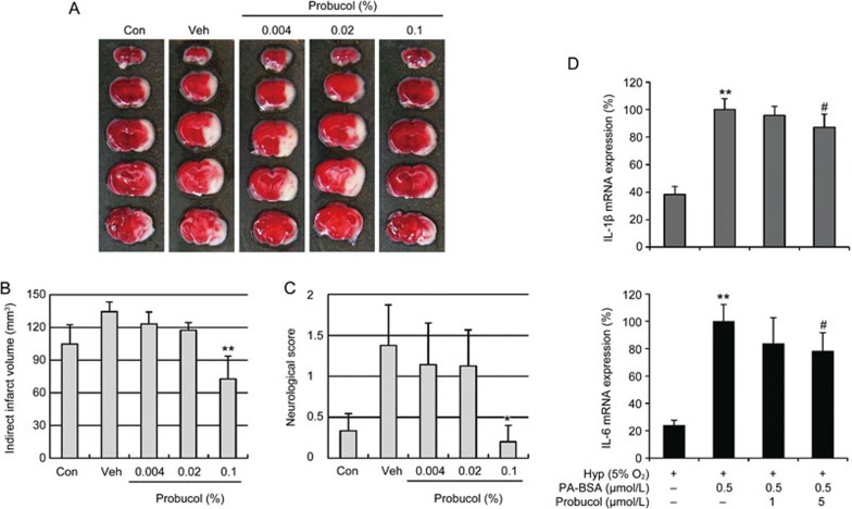
Effects of probucol on the infarct volumes and functional outcomes following focal cerebral ischemia in hyperlipidemic mice. (A) Representative topical TTC-stained brains from ApoE KO mice that were fed HFD with or without 0.004%, 0.02%, or 0.1% probucol for 10 weeks. The mice were subjected to 45 min of MCA occlusion followed by 24 h of reperfusion. White indicates the infarct area. (B) Quantification of the infarct volumes 24 h after ischemia (n=7–8). The infarct volumes were calculated by integrating the infarct areas in 2 mm-thick coronal slices. (C) The neurological deficits of each mouse were assessed 24 h after ischemic insult in a blinded fashion (n=7–8). The values are presented as the mean±SEM. *P<0.05, **P<0.01 vs age- and diet-matched ApoE KO mice (Veh, vehicle). (D) BV2 microglial cells were exposed to in vitro hyperlipidemic conditions (0.5 μmol/L palmitic acid-BSA) with hypoxia (5% O2). Palmitic acid (PA)-BSA treated cells were incubated for 3 h, moved to hypoxic conditions and further incubated for 16 h. We observed the IL-1β and IL-6 mRNA expression with real-time PCR. The PA-BSA-induced upregulations of IL-1β and IL-6 were decreased by probucol (n=6). **P<0.01 vs hypoxia group (Hyp); #P<0.05 vs PA-BSA with hyp group (Veh).

