
FIGURE 1.
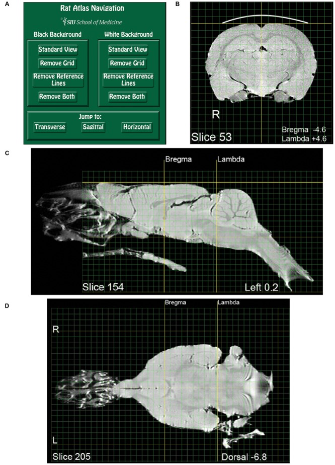
Pages from the magnetic resonance images (MRI) rat atlas. (A) Depiction of the atlas home page with control buttons for navigation and selection of display settings. (B) An exemplary transverse (i.e., coronal) slice located midway between Bregma and Lambda. (C) An exemplary sagittal slice located 0.2 mm left-lateral of midline. Note that the olfactory epithelium and the bony and soft palate are included in the image. (D) An exemplary horizontal slice located 6.8 mm ventral to the dorsal-most surface of the brain [indicated by the gold horizontal reference line in (C)].