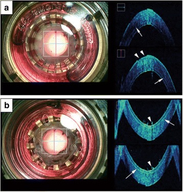
Fig. 2.

Intraoperative SD-OCT image of a donor cornea for precut Descemet’s stripping automated endothelial keratoplasty (DSAEK) observed through a viewing chamber. a Representative microscope image (surgeon’s view) observed from the anterior (left). Normal corneal curvature and stromal texture together with high reflectivity of the epithelium, microkeratome cut line (arrows), endothelium cell layer and epithelial defect (arrowheads) were observed by intraoperative OCT (right). b Microscopic image observed from the posterior cornea (left) and intraoperative OCT image (right). Characteristic microkeratome cut line (arrows) together with debris (arrowheads) observed on the endothelium cell surface was observed (left)
