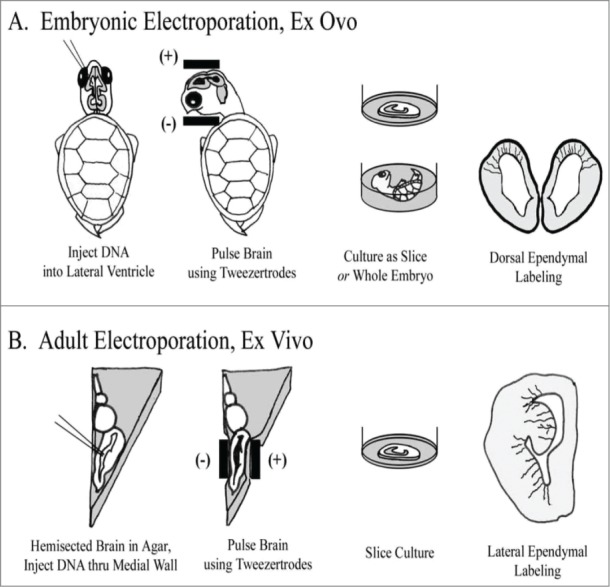
Figure 1.

Individual RG cells were labeled using electroporation. (A) In embryonic animals, ex ovo electroporation was used. Subsequently, brains were either cultured as coronally sectioned slices or cultured in the whole embryo. Electrodes were applied following a dorso-ventral orientation to label cells in the dorsal cortex. (B) In adult animals, the brain was removed, hemisected and embedded in agarose. Electrode positioning following a medio-lateral orientation to label cells in the DVR and striatum. After electroporation, brains were cut into slices and organotypic slice cultures prepared.
