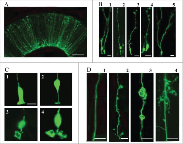
Figure 2.

Electroporation of the embryonic turtle telencephalon with an EGFP-expressing plasmid labels many cells with RG morphology. (A) At 24 hours post-electroporation of a stage 20 embryo, an array of EGFP+ cells in the dorsal cortex show RG morphology. (B) At higher magnification a variety of morphologies are seen. RG cells in different phases of the cell cycle are apparent (B1, B2). Small expansions are often present along the length of the pial process (B3, B4). A smaller number of EGFP+ cells in the VZ do not appear to have a pial process. These may be newly generated neurons, intermediate progenitor cells, or RG cells. At later stages (stage 23), cells are found with bifurcated or branched pial processes, and ‘hairy’ or more lamellate fibers (B5). (C) A variety of ventricular contacting processes are seen. Some processes are typical for embryonic RG cells seen in rodents (C1, C2) while others are more complex (C3, C4). (D) RG cells exhibit different heterogeneous morphologies. D1, RG cells in younger animals often have a smooth pial process. In many cases expansions bud off of the process (D2) or in line with the process (D3). As development proceeds RG cell pial fibers exhibit more mature ‘lamellate’ fibers (D4). Scale bars: A, 50 μm; B, 10 μm; C,D, 10 μm.
