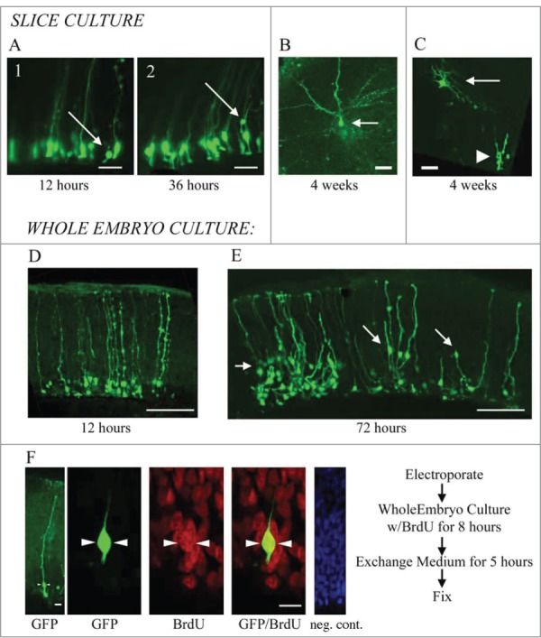
Figure 3.

RG cells migrate, exhibit cellular movements, and proliferate in organotypic slice cultures and in whole embryo cultures. (A) RG cells in the embryonic turtle telencephalon labeled through electroporation. Shown are 2 time points, 12 hours and 36 hours. Electroporated cells were adjacent to the ventricle at early time points (A1), but at later time points, some RG cell bodies had translocated away from the ventricular surface (A2). (B and C) Slice cultures allowed to incubate for up to 4 weeks contained cells with mature neuronal morphologies. In C, the neuron (arrow) is located just superficial to the proliferating area of ependymal cells (arrowhead). (D and E) Representative examples of dorsal cortex that were incubated in whole embryos cultures for 12 hours (D) and 3 d (E). At 12 hours, RG cell bodies were close to the ventricle. At 72 hours, many electroporated cells had migrated or translocated into the cortex (arrows in E). (F) Whole embryos cultured with a pulse of BrdU. Left panel shows a single electroporated RG cell (green). The second panel is a higher power image of the same cell. BrdU staining revealed many mitotic cells (red), including GFP+ RG cells that were BrdU+ (green, arrowheads). The right panel is a negative control BrdU staining. DAPI stain (blue) labels all nuclei. Scale bars: A, B, C, 20 μm; D, E, 100 μm; F, 5 μm.
