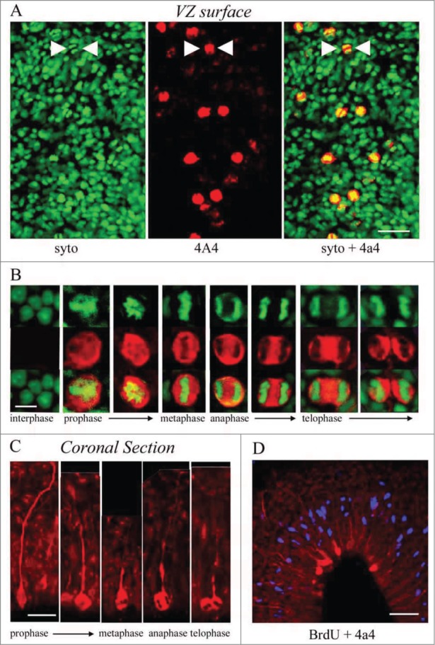
Figure 4.

M-phase cells in the embryonic turtle VZ express the RG lineage marker phosphorylated vimentin (4A4) and reveal RG cell morphology. (A) En face cortical slab stained with syto-11 (green) and 4A4 (red). All M-phase cells at the ventricular surface express 4A4. (B) 4A4 robustly labels M-phase cells throughout mitosis. (C) In coronal sections 4A4 labeled mitotic cells lining the ventricle that possessed pial fibers coursing out through the parenchyma. Pial fibers were most robust in prophase, but a thin process remained throughout division. (D) A brief BrdU (blue) pulse shows that S-phase cells are found in an abventricular position at the top of the VZ while 4A4+ cells mitotic cells were located at the ventricular surface, indicating interkinetic nuclear migration in the developing turtle telencephalon. Scale bars: A, 20 μm; B, 5 μm; C, 10 μm; D, 20 μm.
