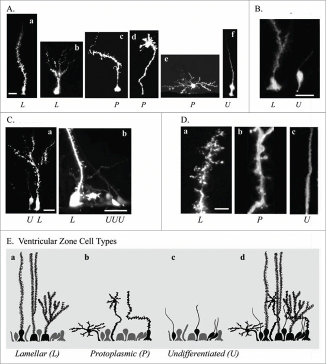
Figure 6.
Electroporation of the adult turtle telencephalon reveals heterogeneous RG cells that we grouped into 3 categories distinguishable by their pial fiber morphology. Lamellate RG cells (L, Aa, Ab); Protoplasmic RG cells (P, Ac−Ae); and Undifferentiated RG cells (U, Af). (B) Further examples of the 3 cell types identified by letter under each image. (C) Lamellate RG cells have pial fibers possessing ‘hairy’ fine extensions, and a pial fiber that in some cases had multiple branches within the parenchyma. (D) Comparison of the pial fiber of the 3 cell types in higher magnification images. Protoplasmic fibers had many smooth expansions. Cell bodies were located at the ventricle and away from the ventricle. Protoplasmic RG cells had the most diverse cellular morphologies. Undifferentiated fiber types were smooth and traceable through the pyramidal cell layer and for several hundred micrometers into the parenchyma. They arose from smaller cell bodies most frequently found close to the ventricle (B and C). (E) Schematic showing the 3 classes of RG cells and their overlapping distribution (Ed). We hypothesize that undifferentiated RG cells retain the capacity for proliferation. Scale bars: A, B, C, 10 μm; D, 3 μm.

