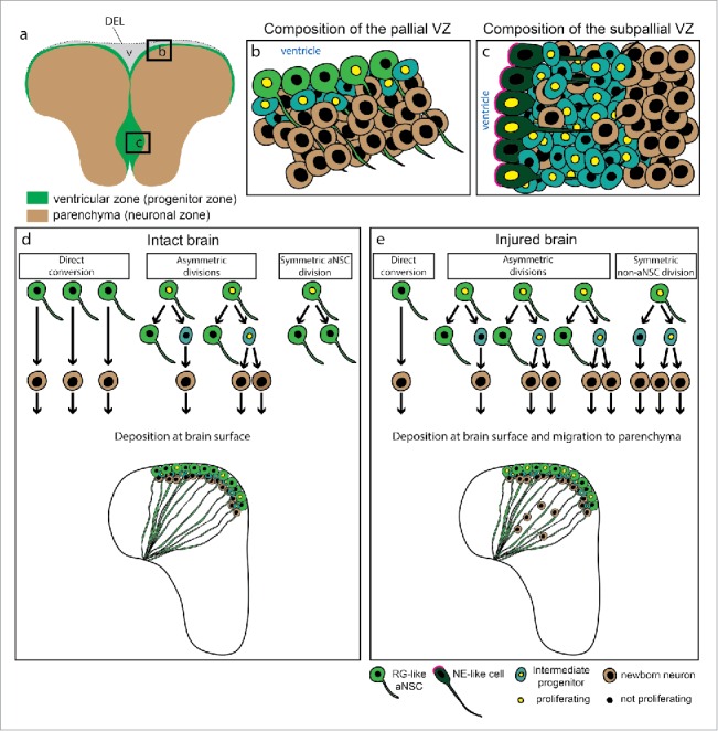
Figure 1.

Neurogenic niches in the adult zebrafish telencephalon and behavior of aNSCs in the pallium. (a) Representation of a coronal section through the adult zebrafish telencephalon illustrating the ventricular zone (green), containing aNSCs and progenitors, and the parenchyma (brown), mostly composed of neurons. The two hemispheres are linked by a dorsal ependymal lining (DEL) that closes the ventricle (v). (b) and (c) Scheme of the dorsal (b) and ventral (c) ventricular zones, composed of different progenitor types: RG-like aNSCs, non-glial intermediate progenitors and neuro-epithelial-like cells. (d) and (e) Modes of cell division and neurogenesis in the intact (d) and injured (e) zebrafish telencephalon, assessed by live imaging.10 In the intact brain (d) the newborn neurons are deposited immediately adjacent to the progenitor cells. After injury (e) there is an increase in proliferating aNSCs at the VZ, a change in their mode of cell division and the migration of progeny to the injured parenchyma, where the newborn neurons contribute to tissue regeneration. The dashed circle marks the region where the lesion was. Abbreviations: DEL-dorsal ependymal lining; NE-neurepithelial; RG-radial glia; VZ-ventricular zone.
