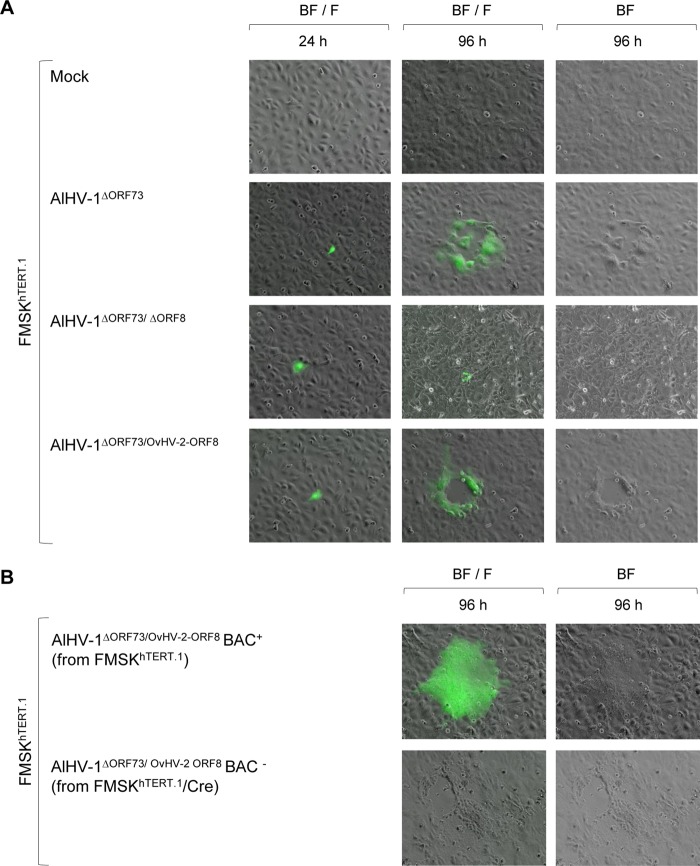
FIG 2 .
Plaque formation and viral replication in cell culture. (A) Representative fluorescence microscopy images of FMSKhTERT.1 cells transfected with AlHV-1 BAC DNA from different constructs at 24 and 96 h posttransfection. Nontransfected cells (mock) were used as a control. Virus spreading and cytopathic effect are indicated by the formation of plaques. Green fluorescence indicates expression of green fluorescent protein encoded by the BAC cassette. BF, bright field; F, fluorescence with a fluorescein isothiocyanate (FITC) filter. Magnification, ×10. (B) Representative images of FMSKhTERT.1 cells infected with AlHV-1ΔORF73/OvHV-2-ORF8 reconstituted from FMSKhTERT.1 (BAC+, BAC cassette intact) or FMSKhTERT.1/Cre cells (BAC−, BAC cassette excised) at 96 h postinfection.

