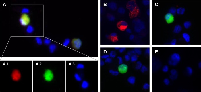
FIG 3 .
Reactivity of OvHV-2 gB-specific antibodies to the AlHV-1ΔORF73/OvHV-2-ORF8 virus. Representative fluorescence microscopy images of FMSKhTERT.1 cells harvested at 24 h posttransfection with AlHV-1ΔORF73/OvHV-2-ORF8 (A and C), pOvHV-2 ORF8 (B), or AlHV-1ΔORF73/ΔORF8 (D) DNA and untransfected cells (E). Cells were treated with OvHV-2 gB hyperimmune mouse serum (A, B, D, and E) or preimmune serum (C) as a primary antibody and an anti-mouse IgG conjugated to Alexa Fluor 568 as a secondary antibody. Slides were mounted with SlowFade Gold Antifade Mountant with DAPI and examined using fluorescence microscopy. Individual images from the indicated area of merged image A are shown in A.1, A.2, and A.3. Red fluorescence indicates reactivity of serum antibodies with gB, green fluorescence indicates expression of green fluorescent protein encoded by the BAC cassette, and cell nuclei are stained blue. Magnification, ×20.

