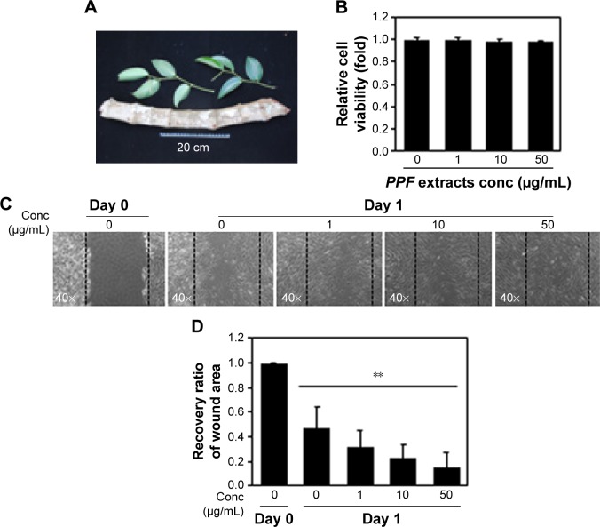
Figure 1.
Cell viability and wound-healing assays.
Notes: PPF leaves and stem picture. (A) PPF extract applied at 1 and 10 μg/mL showed no cytotoxicity in the EXCyto assay using NHDF cells. (B) The cell wound-healing assay was imaged 24 hours after scratching the cell monolayer and treating with PPF extract at the indicated doses. (C) Once cells reached confluence, a single wound was made in the center of the monolayer using a 10 μl pipette tip and the cells were treated with the indicated concentration of PPF extracts. After a 24-hour incubation, a photograph was taken at ×10 magnification under a microscope. (D) The photographic images from (C) in a dose-series were analyzed for gap area. The results are presented as the mean ± standard deviation of three independent experiments. **P<0.01.
Abbreviations: Conc, concentration; NHDF, normal human dermal fibroblast; PPF, Piper cambodianum P. Fourn.

