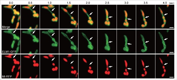
Figure 1.
Time-course observation of the bridging of two mitochondria by a mitochondrial outer-membrane protrusion (MOP). These are representative images from a movie (Supplemental Movie 1) that was recorded at 10 frames per second. The movie was obtained by VIAFM (Variable incidence angle fluorescent microscopy20) using leaf epidermal cells from 10-day-old transgenic Arabidopsis plants expressing ELM1-GFP and Mt-RFP. The arrows indicate a MOP bridge, and the bridge was stretched for a few seconds. The time stamp is in half seconds. Bars = 1 µm.

