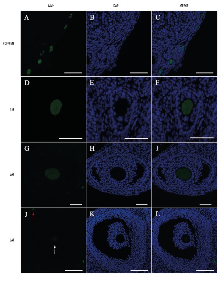
Figure 1.
Localization of MVH protein in the ovary by immunofluorescence. Green: Anti-MVH staining. Blue: DAPI-stained nuclei. MVH is expressed in the cytoplasm of oocytes in (A–C) primordial follicles (PDF) and primary follicles (PMF), (D–F) secondary follicles (SEF), (G–I) small antral follicles (SAF), and (J–L) large antral follicles (LAF). Arrows in (J) display a very weak MVH expression in large antral follicles (white arrow) compared with a strong one in primordial follicles (red arrow). Scale bars in (A–I): 50 μm, in (J–L): 100 μm.

