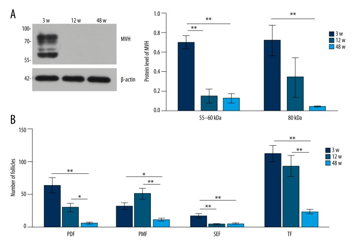
Figure 3.
Expression pattern of MVH protein in 3-, 12-, and 48-week-old female mice. Western blot for MVH in the bone marrow in 3-, 12-, and 48-week-old female mice. N=10 each. (B) Western blot for MVH in the ovary in 3-, 12-, and 48-week-old female mice. N=4 each. The numbers on the left in (A, B) are the size standards of molecular weight (in kDa).

