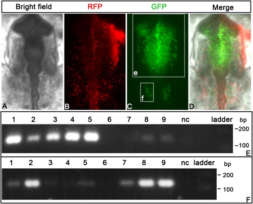
Figure 4.

Detection of enhancers in neural tube and neural crest. A–D: Embryos were electroporated with a mixture of barcoded plasmids containing FoxD3‐NC1 and ‐NC2, Sox10E, Sox2‐N2, Sox2‐N4, mSix1‐21 1x/2x/4x enhancers, empty vector, and β‐actin promoter‐driven RFP at HH6/7. At HH10, enhancer activity (EGFP) is seen in the neural tube, the neural crest, and otic placode (C, D), while RFP expression is widespread. The white rectangles e and f indicate the regions dissected for assaying enhancers. E: Positive enhancers in the head region are detected by the 129‐bp bands (FoxD3‐NC1: lane 1, Sox10E: lane 2, Sox2‐N2: lane 3, Sox2‐N4: lane 4, FoxD3‐NC2: lane 5, Six1‐21‐2x: lane 8, Six1‐21‐4x: lane 9). Lane 6, 7, and 10 show Sox2‐N3, Six1‐21‐1x, and the empty vector, respectively. F: Otic enhancers are captured by the assay. Sox10E (lane 2), Six1‐21‐1x/2x/4x (lanes 7–9) produce the specific band around 129 bp. Lane1 (FoxD3‐NC1) and lane 5 (FoxD3‐NC2) show weak signal probably resulting from neural crest contamination. The neural tube enhancers (Sox2‐N2: lane 3, Sox2‐N4: lane 4, Sox2‐N3: lane 6) are negative in the otic placode.
